
A single chip with multiple talents
The Medipix chips developed at CERN are being used in a variety of fields: from medicine to education and back to high-tech engineering. The scene is set for a bright future for this versatile technology.
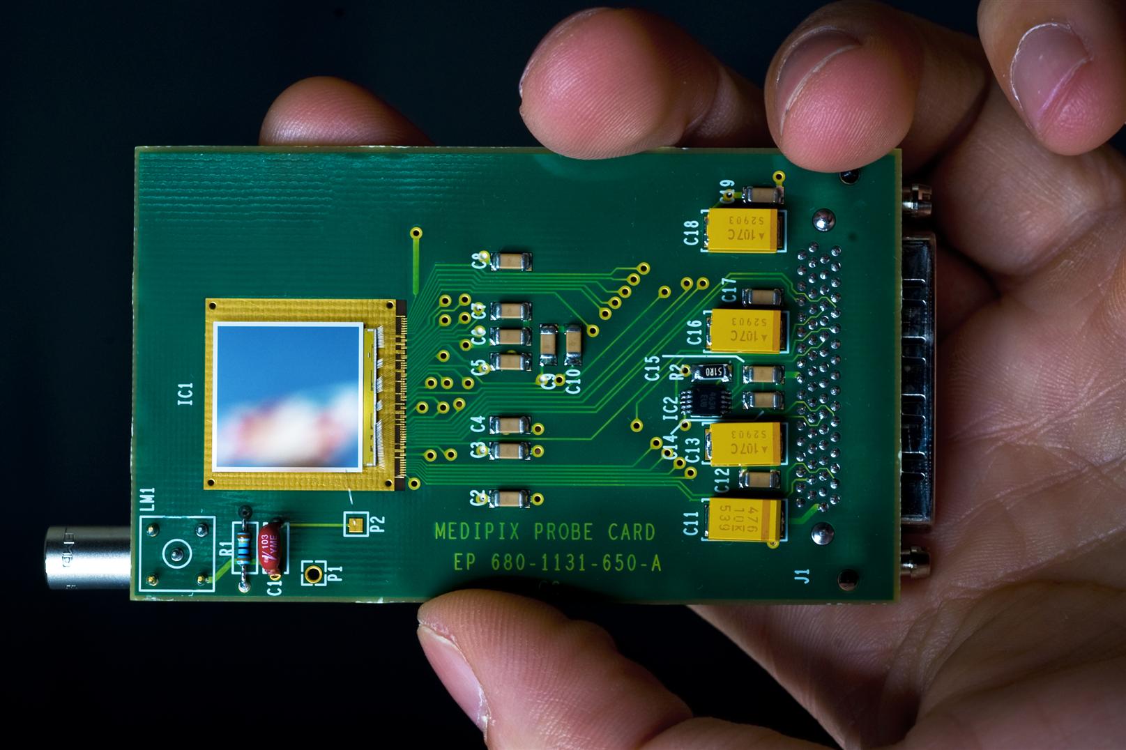
The Medipix chip.
Radiography and computed tomography (CT) use X-ray photons to study the human body. The different energies of the photons in the beam can be thought of as the colours of the X-ray spectrum. This is why the use of Medipix3 chips in such diagnostic techniques is referred to as colour X-ray imaging. The images become clearer, and more accurate and it becomes easier for doctors to distinguish the different tissues (read the interview below). “Our initial goal back in the mid 90’s was to develop a single photon-counting pixel detector with a very high signal-to-noise ratio”, says Michael Campbell, spokesman for the Medipix3 collaboration. “The Medipix chips we have developed (Medipix1, Medipix2, Timepix and now Medipix3) have found applications in medicine, but also in accelerators and even in education”.
Indeed, the chips are suited to a number of different applications. The Medipix2 chip has been used by the UA9 experiment to monitor the beam halo in the collimation region of the SPS in an extremely accurate way.
More surprisingly, the Timepix chip which is derived from Medipix2 is being used in a very ambitious educational project led by the Simon Langton Grammar School in the UK. Teams of high-school students are using the Timepix chips as cosmic ray detectors. The Langton Ultimate Cosmic ray Intensity Detector (LUCID) will fly on a Surrey Satellite Technology Limited satellite in 2011 and will monitor cosmic rays from space. At the same time, students from different schools are joining forces and, by simply connecting the Timepix chip to their computers through a USB interface, are creating a network of cosmic ray detectors on Earth. The Bulletin will cover this topic in more detail in the next issue.
Medipix seems to have taken a sort of round-trip from high-energy physics to medicine and back. The design team is starting work on a new chip which will act as a prototype for the upgrade of the LHCb vertex locator. This follows on the needs of applications in beta and gamma radiography of biological samples, materials analysis (together with an industrial partner), monitoring of nuclear power plant decommissioning, electron microscopy, and adaptive optics relevant to large ground-base telescopes…
Interview with Dr Anthony Butler, Radiologist at the University of Otago Christchurch and the University of Canterbury (New Zealand) and head radiology researcher on the Medipix project.
CERN Bulletin: How do you think X-ray colour technology will benefit patients?
There are several ways that the technology will benefit patients.
1) A more efficient use of current radiographic contrast agents, or radiographic pharmaceuticals.
Currently in medicine we use radiographic pharmaceuticals that show up on X-ray imaging. These are seen as "dense" or "bright" areas on current X-ray systems. On colour imaging systems, like Medipix3, these agents have their own X-ray colour.
This allows a few things:
- potentially reduced dose of the pharmaceutical. This is good for the patient because the pharmaceuticals are toxic to kidneys, especially in diabetes and elderly people.
- ability to distinguish the pharmaceutical from other bright objects. This is important in things like blood vessel disease where either bone or calcium in the vessel make identification of the pharmaceutical difficult.
- ability to have several pharmaceuticals at the same time. This is important, as often the radiologists would like to give pharmaceuticals at different times and separately evaluate their perfusion through he body. For instance, for renal imaging it’s good to give one contrast agent 15 minutes before the scan and a second agent 30 seconds before the scan. On current systems this requires multiple scans. With colour imaging several different pharmaceuticals can be given at the different times, then separately identified based on their X-ray colour.
A transverse section through a mouse’s chest in which the x-ray colour of different radiographic pharmaceuticals is demonstrated. Iodine based compounds are shown as red in the blood vessels of the lungs and heart. Barium based pharmaceuticals are shown as yellow in the air spaces of the lungs. Background tissues, such as bone, are shown as white.
Several imaging artefacts occur because the quality or colour of the X-ray changes as it passes through the body. Devices like Medipix3 allow this to be measured and therefore corrected for, thus improving image quality.
3) New pharmacological contrast agents.
New contrast agents are being developed that have specific X-ray colours. For example, groups are working on labelling cancer cells with pharmaceuticals with specific colours. Other groups are working on pharmaceuticals that label enzymes (eg. ACE) in the blood pressure control system.
4) Improved tissue discrimination.
Often in a thin tissue there are two objects that look very similar on black and white imaging. Examples include calcium and iron within diseased blood vessel walls. People are using Medipix to try to better separate these two elements. Their goal is to have better prediction of the stability of the diseased vessel wall, thus allowing better treatments for conditions like heart disease and stroke.