
Crystals against cancer
This is a remarkable example of direct technology transfer from particle physics to medicine. Clinical trials have begun in Portugal on a new medical imaging system for the diagnosis of breast cancer, which uses positron emission tomography (PET). The system, developed by a Portuguese consortium in collaboration with CERN and laboratories participating in the Crystal Clear collaboration, will detect even the smallest tumours and thus help avoid unnecessary biopsies.

Antimatter is a useful commodity…and not only for making bestsellers and Hollywood blockbusters! A groundbreaking new positron emission tomography (PET) device has just been commissioned for the first time in Europe. If trials are successful, it promises to be an important step forward in the diagnosis of breast cancer. In February, the PET-Mammography consortium of eight Portuguese institutes, including the national particle physics laboratory LIP, Lisbon’s Garcia de Orta Hospital and the Porto Institute of Oncology, commissioned a prototype PET device dedicated to breast imaging, ahead of clinical trials. The device was developed over the last five years in the framework of the Crystal Clear collaboration. The collaboration, whose spokesman is Stefaan Tavernier of Brussels’ Vrije Universiteit, has been in the vanguard of crystal work for particle physics and its spin-off applications since 1990.
Until now, positron emission tomography (PET) devices were conceived as large machines capable of "photographing" the whole body (see box). But a few years ago, the members of the Crystal Clear collaboration came up with the idea of developing dedicated PET devices with the aim of achieving better results. It was quickly established that breast imaging lends itself well to this technique, as the breast is an "external" organ that can easily be "placed" inside such a device. Moreover, the field is crying out for an improved breast imaging device, for although the spatial resolution of conventional X-ray mammograms is excellent, the results are often inconclusive. According to CMS physicist and initiator of the Crystal Clear collaboration, Paul Lecoq, "60% of positive X-ray results, i.e. those indicating a potential problem, turn out not to be cancers." Mammogram results can be particularly misleading in the case of younger women, who have dense mammary tissue, women who have had plastic surgery and women undergoing treatment for the menopause. In such cases doctors tend to prescribe a biopsy which, although a relatively light procedure, is still both invasive and costly.
For these patients, the new device, known as ClearPEM (PEM standing for positron-emission mammogram), is a powerful diagnostic tool that can help avoid many unnecessary biopsies. "This is great news for millions of patients across Europe," says Paul Lecoq. The advantages with respect to conventional PET devices are evident. ClearPEM is more sensitive than PET, detecting 5 to 10 times more emitted radiation for the same injected dose (see box). This higher sensitivity makes it possible to detect smaller tumours, which emit fewer gamma rays - ClearPEM can identify tumours 2 to 3 mm in diameter, compared with 10 to 15 mm for a "full-body" PET. The examination also takes less time. "It can be done in 5 minutes instead of the 25 needed for a conventional PET scan," explains Joao Varela, a CMS physicist who also leads the Portuguese collaboration in ClearPEM. Shorter analysis time also means a smaller injected radioactive dose.
So the challenge was to build a PET device compact enough to be applied to a single organ. Developments began in 2003, using techniques employed for the CMS electromagnetic calorimeter. "ClearPEM works in exactly the same way as a crystal electromagnetic calorimeter. The only difference is one of scale," Varela adds. In this respect, ClearPEM is a great example of the direct transfer of technology from particle physics to medicine.
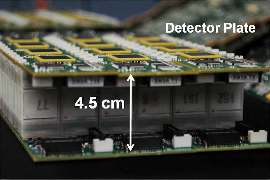
The device consists of two 16 x 18 cm plates constituted by a matrix of crystals, inserted together with the associated electronics into an automated rack. During the scan, the device rotates about the breast. Like the CMS calorimeter, ClearPEM is endowed with crystals that scintillate as the high-energy photons (gamma rays) emitted by the body pass through them. The 6,000 crystals in ClearPEM are far more sensitive than those in CMS, however, as they need to detect much weaker signals. And they are obviously much smaller as the energies of the gamma rays to be detected are approximately 100,000 times weaker. The crystals were characterised at CERN using a device similar to the one used to characterise the CMS crystals.
One of the keys to achieving a compact device was the use of avalanche photo-diodes (APD), which transform the light signal into an electrical one. These very compact silicon cubes, affixed to the ends of the crystals, were developed for CMS. "We also had to develop very compact, very low-background electronics, as well as read-out electronics based on the CMS trigger system," adds Varela, who is none other than the person responsible for the CMS trigger system.
The prototype is now being used to perform 100-200 clinical trials at the Porto Institute of Oncology. Depending on the results obtained, the medical community may decide to put such a device into full service. Meanwhile, the members of the Crystal Clear collaboration have other projects up their sleeves, notably in the framework of the CERIMED project. CERIMED (European Centre for Research into Medical Imaging), a project being set up in Marseilles under the impetus of its promoter Paul Lecoq, is in the process of building a PET device coupled to an ultra-sound system. This new device, known as ClearPEM sonic, is due to be installed in Marseille’s La Timone Hospital by the end of 2009. The marriage of PET and ultra-sound technologies promises another leap forward in breast imaging.
How does a PET scanner work?
Positron emission tomography (PET) is a medical imaging technique allowing 3-D measurements of an organ’s metabolic activity. The patient is injected with a sugar that is marked with a radioactive substance. Cancerous cells, which are more active than healthy ones, will more readily absorb this special sugar, which is not metabolised by cells. When the radioactive marker, called Fluor 18, decays, it emits positrons that in turn annihilate and emit gamma rays (photons) in opposite directions at a known energy (511 keV). The crystals of the PET device, arranged on two plates positioned either side of the body or the organ, detect these photon pairs, in the same way as the CMS crystal calorimeter will detect the two gamma rays produced by the decay of the Higgs boson. By reconstituting all the coinciding events (the two photons emitted at the same time in opposite directions), the device can reconstruct an image and observe the metabolism of the cells. This is why we refer to functional imagery, as opposed to X-ray imaging techniques, for example, which provide anatomical imagery.